Cavovarus Foot Reconstruction Surgery
Cavovarus Foot Reconstruction Surgery Information Sheet
What is Cavovarus Foot Reconstruction Surgery?
Cavovarus foot reconstruction surgery is a procedure to correct a deformity where the arch of the foot is abnormally high (cavus), and the heel turns inward (varus). This structural imbalance places excessive pressure on the outer part of the foot and ankle, often causing pain, frequent sprains, walking difficulties, and instability. The procedure is usually performed by an experienced Orthopaedic Surgeon in Adelaide to restore balance, improve mobility, and prevent long-term complications.
The reconstruction aims to restore normal foot alignment by realigning bones, balancing tendons, and lengthening tight tissues. The specific surgical approach depends on the severity of the deformity and the underlying cause, which may include neurological conditions, previous injury, or congenital abnormalities.
Who is Suitable for Cavovarus Foot Reconstruction Surgery?
Not every patient with a cavovarus foot requires surgery. However, reconstruction may be appropriate for the following individuals:
- Persistent Symptoms: People who experience chronic pain in the ball of their foot or on the outside of their foot, ankle instability, frequent ankle sprains, or 5th metatarsal stress fractures that do not respond to conservative treatments like orthotics or physiotherapy.
- Progressive Deformity: Individuals whose foot structure continues to deteriorate over time, affecting balance, gait, or the ability to walk.
- Neuromuscular Conditions: Patients with neurological disorders such as Charcot-Marie-Tooth disease, polio, spinal cord injury, or stroke who develop muscle imbalance leading to foot deformity.
- Mechanical Instability: Those with severe foot misalignment that causes difficulty wearing normal shoes, increased risk of falls, or uneven wear patterns on footwear.
- Failed Non-Surgical Treatment: Individuals who have tried braces, shoe inserts, physiotherapy, and medication without improvement.
Benefits of Cavovarus Foot Reconstruction Surgery
Reconstructive surgery aims to restore both function and comfort. Key benefits include:
- Improved Foot Alignment: Correcting the arch and heel position restores more normal walking patterns and gait mechanics.
- Reduced Pain: Offloading pressure from the outer edge of the foot relieves pain, especially in the lateral foot, ankle, and forefoot.
- Greater Stability: Rebalancing tendons and correcting bone alignment helps prevent ankle sprains and falls.
- Better Mobility: Improved weight distribution and joint function make it easier to walk, run, or stand for longer periods.
- Improved Footwear Fit: Patients can wear standard shoes more comfortably without requiring custom bracing or orthotics.
- Prevention of Further Damage: Correcting the deformity can prevent future joint arthritis, tendon rupture, and the progression of deformity.
Types of Cavovarus Foot Reconstruction Surgery
Cavovarus reconstruction is highly individualised and may involve multiple techniques performed during a single operation. Common components include:
- Soft Tissue Procedures:
- Plantar Fascia Release: Cutting tight bands on the sole of the foot to lower the arch and reduce rigidity.
- Tendon Transfers: Often, the peroneus longus tendon is transferred to the peroneus brevis to balance muscle forces and stabilise the ankle. Tibialis posterior is often a deforming force, and is transferred to help lift the foot up
- Achilles Tendon Lengthening or Gastrocnemius Recession: Used if calf muscle tightness is contributing to the deformity.
- Bone Procedures:
- Calcaneal Osteotomy: The heel bone is cut and shifted to correct varus heel alignment.
- Dorsiflexion Osteotomy of the First Metatarsal: A wedge of bone is removed to lower the forefoot and realign the arch.
- Midfoot Osteotomies: Addresses fixed deformities in the midfoot, often in combination with other corrections.
- Circular frame: For severe deformities, an external device can gradually change the foot shape over time for a more accurate result
- Joint Fusions (Arthrodesis):
- Reserved for severe deformities or cases involving arthritis.
- May include fusion of joints in the hindfoot or midfoot to provide a stable, pain-free platform.
- Neuromuscular Adjuncts:
- If a neurological condition is present, procedures may also address muscle tone or spasticity, such as selective tendon lengthening or balancing opposing muscle forces.
Alternative Options to Cavovarus Foot Reconstruction Surgery
Surgery is not always the first step in treating cavovarus foot. Many people benefit from non-surgical treatments, especially in the early stages or if the deformity is mild and flexible. The main alternatives include:
- Custom Orthotics: Specially made shoe inserts help to support the arch, reduce pressure on the outer foot, and improve balance.
- Ankle Bracing: Devices such as ankle-foot orthoses (AFOs) can provide stability and reduce the risk of ankle sprains.
- Footwear Modifications: Supportive shoes with lateral wedges, cushioned insoles, and high-top designs help to control foot position and protect the ankle.
- Physiotherapy: Targeted exercises can strengthen weak muscles, improve balance, and stretch tight tendons, thereby contributing to the correction of deformity.
- Activity Modification: Avoiding high-impact or unstable activities can reduce pain and prevent further injury.
- Medications: Anti-inflammatory medicines or topical treatments may help reduce discomfort in patients with joint pain or muscle strain.
- Botox Injections: In select patients with neuromuscular causes, Botox may be used to temporarily reduce spasticity or muscle overactivity.
Preparation Before a Cavovarus Foot Reconstruction Surgery
Proper preparation can help reduce complications and support a smoother recovery:
- Medical Evaluation: Have blood tests, imaging (X-rays, CT, or MRI), and general health checks to confirm surgical suitability.
- Medication Review: Inform Dr Graff about all medications. Some may need adjusting before surgery.
- Stop Smoking: Quit smoking at least a few weeks before surgery to support healing.
- Arrange Support: Plan for time off work, organise help at home, and get mobility aids like crutches or a walker.
- Prepare Your Home: Set up a safe recovery space with easy access to essential items and bathroom safety supports.
- Understand the Plan: Discuss the procedure, risks, implants, and recovery expectations with Dr Graff.
- Check Costs: If going private, confirm all fees and what your insurance covers.
- Pre-Book Physio: You will need physiotherapy soon after surgery—booking early can help avoid delays.
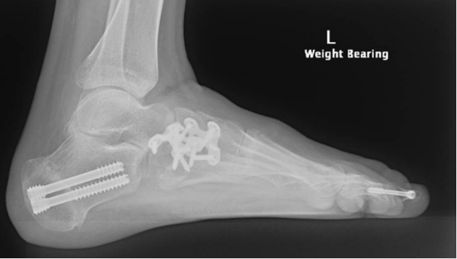
Cavovarus Foot Reconstruction Surgery Procedure
The surgery is not a single procedure, but rather a collection of surgical techniques tailored to each patient’s specific condition. Common procedures include a combination of:
- Osteotomy: Realigning or reshaping the bones to help restore the lower arch and shift the heel
- Tendon Transfer: Tibialis posterior tendon transfer from the inside to the top of the foot
- Soft tissue procedures: releasing the tight plantar fascia, tendoachilles lengthening.
- Bone Graft: Sometimes, bone graft from another part of the body (often the pelvic bone near the hip) is used.
- X-rays are used in theatre to make sure the bones are aligned correctly, and a tourniquet is placed around the thigh to help with bleeding.
- The surgery can take 90 to 120 minutes.
The Hospital Stay
- You wake up with a half plaster (backslab) or a boot
- Your foot will be elevated overnight, and you will have antibiotics through a drip
- You will either be given blood-thinning medication to help prevent DVT and vitamin C to help with wound healing
- You will stay in the hospital for 2-5 days with antibiotics, blood thinners, regular paracetamol, regular laxatives, regular vitamin C and stronger pain killers to take if and when required
- You will only be allowed to touch your foot to the ground for 6 weeks
- Depending on your balance and strength, you may need rehabilitation post-operatively
- Buying a second-hand knee scooter (you can search online) and practising at home before surgery can be helpful; please bring it with you to the hospital. It is easier to use a knee scooter than crutches
When You Go Home
- You will need medications for pain relief; regular paracetamol (2 tablets four times a day) is recommended, as well as strong pain killers, especially at night before bed. These can have side effects of drowsiness, nausea and constipation, and other tablets to help with these side effects may be required.
- You will need to take blood thinners and vitamin C as prescribed
- You will need a shower chair and bags to keep the plaster dry
- You will need to attend your post op appointment in 2-3 weeks, where the wounds will be checked and your plaster changed to a waterproof cast if your wounds are healed
- After this, you will be able to shower with a shower chair and get the plaster wet
Cavovarus Foot Reconstruction Surgery Rehabilitation
All patients are different. These timelines are only guides; some patients may progress more quickly or slowly than others.
0 - 2 Weeks
- You will be in a backslab or moonboot for 2-3 weeks
- You will only be allowed to touch your foot to the ground for balance. Please keep your foot elevated and out of bed for the toilet only
- You will need to bag the leg for showers
- Pain relief: Please take regular paracetamol with meals and before bed; stronger pain killers are often needed, especially before bed
- Please take blood thinners and vitamin C as prescribed
2 - 3 Weeks
- Post op appointment: dressings are changed, and an X-ray is taken
- You will then go into a full cast or stay in the moonboot full time for another 3-4 weeks
- You will still need to elevate the foot at rest
- You can start static strengthening leg lifts with physiotherapy
6 Weeks
- You will have a check X-ray
- If you have a cast, it will be removed, and you can start weight bearing in a boot for 6 weeks
- You can continue isometric tibialis posterior strengthening and retraining and active range of movement with physiotherapy
12 Weeks
- You will have another check X-ray.
- You can start weight bearing without the boot if you can fit into normal shoes You can start swimming and cycling
- You can commence eccentric tibialis posterior strengthening with physio
When can I drive?
- Left foot 3 weeks (if driving an automatic car)
- Right foot 8 -12 weeks
When can I return to work?
- Seated work at 6-8 weeks
- Prolonged standing 6-9 months
- Heavy labour work 12-18 months
Cavovarus Foot Reconstruction Surgery Prognosis
The majority of patients experience significant improvement in pain, stability, and foot alignment. Long-term outcomes are best when surgery is combined with consistent rehabilitation and footwear modifications. Most people return to work and their daily activities, and some can resume sports and hiking, depending on the extent of their pre-existing damage.
Cavovarus Foot Reconstruction Surgery Risks
- Anaesthetic problems
- Nerve injury
- Blood clots
- Infection
- Stiffness
- The osteotomy does not heal (nonunion)
- The osteotomy heals in the wrong position (malunion)
- Ongoing pain
- Arthritis
- The need for further surgery
What if Surgery is Delayed?
Delaying surgery may lead to worsening deformity, chronic pain, ankle instability, and progressive arthritis in the foot and ankle joints. Long-term disability can occur, especially if the underlying cause (e.g., neurological disorder) worsens over time.
Contact Us
If you want more information or have any questions or problems, please contact Dr Graff at admin@christygraff.com or call the rooms at 0493 461 133.