
Talus Osteochondral Defect (OCD)
Talus Osteochondral Defect (OCD) Information Sheet
What is a Talus Osteochondral Defect (OCD)?
A talus osteochondral defect (OCD) is damage to the smooth cartilage of the joint and the underlying bone on the top of the talus bone in the ankle joint. The talus plays a key role in transferring weight between the leg and the foot. When the cartilage is damaged, it can cause pain, swelling, and problems with ankle movement. Sometimes, small fragments of bone and cartilage can break loose into the joint.
Impact of a Talus Osteochondral Defect (OCD) on Anatomy and Health
- Joint mechanics: The talus, like all bones of joints, has a smooth cartilage layer that allows the ankle to move without friction. Damage to this layer disrupts smooth motion.
- Pain and stiffness: This condition can make walking, running, or even standing uncomfortable.
- Long-term risk: Defects in the cartilage can lead to early arthritis, reduced mobility, and chronic ankle problems.
- Swelling: The loss of cartilage can lead to inflammation of the joint, which causes swelling.
Risk Factors for a Talus Osteochondral Defect (OCD)
- Athletes: Particularly those in high-impact sports like football, basketball, and soccer.
- People with ankle injuries: Repeated ankle sprains or fractures increase the risk.
- People with poor ankle alignment: Structural issues in the ankle may contribute to a higher risk.
Causes of a Talus Osteochondral Defect (OCD)
- Trauma: A significant ankle injury, such as a sprain or fracture.
- Repetitive stress: Frequent micro-injuries from sports or physical activity.
- Poor blood supply: In some cases, reduced blood flow to the bone weakens it, leading to collapse and damage.
- Osteochondritis dissecans: This childhood condition can sometimes not heal and lead to an osteochondral defect.
Symptoms of Talus Osteochondral Defect (OCD)
- Ankle pain: Especially after activity or standing for long periods.
- Ankle swelling: Can increase or decrease depending on the amount of activity
- Locking or catching: The ankle may get “stuck” due to loose cartilage fragments.
- Instability: Feeling like the ankle may give way.
- Reduced movement: Stiffness and difficulty flexing or pointing the foot.
Prevention of a Talus Osteochondral Defect (OCD)
- Protect your ankle: Wear supportive shoes, especially during sports
- Strength and balance training: Strong leg and ankle muscles help reduce the risk of injury; physiotherapy can often help after an ankle sprain
- Proper rehabilitation: Fully recover from ankle sprains or fractures before returning to sport.
- Avoid overuse: Allow rest days and avoid repetitive stress on the ankle.
- Early treatment: Seek medical care after significant ankle injuries
Types of a Talus Osteochondral Defect (OCD)
Talus OCD can vary based on the location and nature of the injury:
- Medial lesions: Found on the inside of the talus, usually deeper and cup-shaped. This condition is often caused by repetitive stress or long-standing ankle problems.
- Lateral lesions: Found on the outside of the talus, usually more shallow and wafer-shaped. Often caused by a sudden ankle injury.
- Stable vs. unstable lesions: Stable defects are cracks or damage where the cartilage remains attached. Unstable ones occur when pieces of cartilage and/or bone become loose inside the joint.
Stages of a Talus Osteochondral Defect (OCD)
Doctors use staging systems to describe severity and guide treatment:
- Stage 1: Small area of damage, but the overlying cartilage remains intact
- Stage 2: Cartilage is partially detached but still in place.
- Stage 3: Cartilage and bone are completely detached but not yet displaced.
- Stage 4: Detached fragment has moved into the joint space, causing catching or locking.
Diagnosis of a Talus Osteochondral Defect (OCD)
Diagnosis combines patient history, physical examination, and imaging:
- Clinical examination: Dr Graff will assess for swelling, instability, and range of motion.
- X-rays: May show bone changes but can miss early cartilage damage.
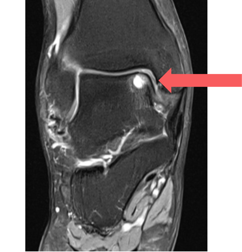
- MRI scan: Best test for detecting cartilage and bone damage, as well as loose fragments.
- CT scan: Provides detailed images of bone defects and helps plan surgery.
Treatment of a Talus Osteochondral Defect (OCD)
Treatment depends on the stage, size, and symptoms:
- Non-surgical options:
- Rest, activity modification, and physiotherapy.
- Anti-inflammatory medications for pain and swelling.
- Bracing or immobilisation to protect the ankle.
- Surgical options:
- Arthroscopic assessment and retrograde drilling: If the cartilage is intact but the bone underneath is damaged, the bone can be drilled through adjacent bone (‘retrograde’) to keep the healthy cartilage intact
- Arthroscopic debridement and microfracture: Damaged tissue is removed using keyhole surgery, and tiny holes are made in the bone to stimulate new cartilage growth; best for small lesions
- Osteochondral grafting (OATS): Healthy bone and cartilage taken from a non-weight-bearing portion of the knee are transplanted to fill the defect.
- Fixation: If the fragment is still intact, it can sometimes be pinned or screwed back in place.
What if a Talus Osteochondral Defect (OCD) is Untreated?
- Chronic pain: Persistent ankle pain during activity or even at rest.
- Joint instability: Increased risk of sprains and further injury.
- Loose fragments: Floating pieces of cartilage and bone may cause locking and catching in the ankle and further damage to the ankle joint
- Arthritis: Progressive damage to the ankle joint leading to early osteoarthritis.
- Reduced quality of life: Difficulty walking, running, or participating in sports and daily activities.
Useful Websites
Contact Us
If you want more information or have any questions or problems, please contact Dr Graff at
admin@christygraff.com or call the rooms at
0493 461 133.